ULTRASOUND


HOW IT WORKS
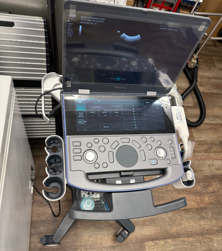
During an ultrasound, high-frequency sound waves capture images of pets’ internal structures. The equipment includes the main ultrasound machine as well as a smaller handheld device. The ultrasound machine emits sound waves, and the handheld device delivers those waves to the patient’s body. The waves bounce off of internal structures, like organs, and return to a sensor in the machine. The machine uses these echoes to instantly produce images we can view on a screen.
Ultrasound is a powerful tool when we need to capture detailed images of soft, fluid-filled organs, including the gallbladder, liver, spleen, and kidneys. Because ultrasound produces images in real-time, we also use this technology to examine the heart as it beats. Ultrasound also helps us detect and diagnose certain types of cancer and monitor pregnancies and fetal health.
![[novatovetclinic.com][493]vet](https://novatovetclinic.com/wp-content/uploads/2023/09/novatovetclinic.com493vet.png)
PET ULTRASOUND IN NOVATO
If your pet needs an ultrasound in Novato or the surrounding areas, we can help. We are proud to provide the advanced diagnostic testing service here at Center Veterinary Clinic and would love to have your pet as a patient. Call now to learn more or schedule an appointment.
WE ARE HERE TO HELP!
Center Veterinary Clinic performs diagnostic ultrasounds for pets in Novato, San Rafael, San Anselmo, Fairfax, Petaluma, and the surrounding areas.

WE ARE HERE TO HELP!
Center Veterinary Clinic performs diagnostic ultrasounds for pets in Novato, San Rafael, San Anselmo, Fairfax, Petaluma, and the surrounding areas.

